Light Microscope
- What Is a Light Microscope?
- Light Microscope Structure
- Light Microscope Applications
- Reasons for Using VHX Series Digital Microscopes
What Is a Light Microscope?

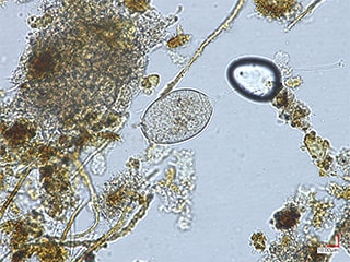
Light microscopes are also called optical microscopes. They use light to perform magnified observation of microscopic targets such as bacteria and blood cells.
Light Microscope Structure
Visual light from the light source is condensed by the condenser lens and projected onto the microscopic, transparent sample. The light transmitted through the sample is once more condensed by a glass lens (the objective lens) for magnified observation.
There are two types of light microscopes: simple light microscopes, which use a single lens, and compound light microscopes, which use multiple sets of lenses.
Simple light microscopes are designed for low magnification. Compound light microscopes are designed for high magnification.
Light Microscope Structure
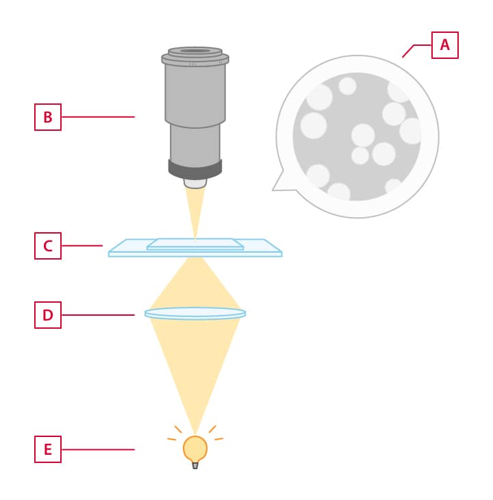
- A
- Brightfield
- B
- Objective
- C
- Specimen
- D
- Condenser lens
- E
- Light source
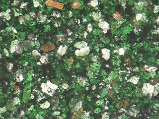
Light Microscope Applications
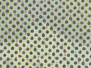
The main observation targets are transparent samples such as bacteria and blood cells. Light microscopes are also known as biological microscopes. They are used to observe organisms.
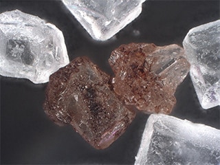
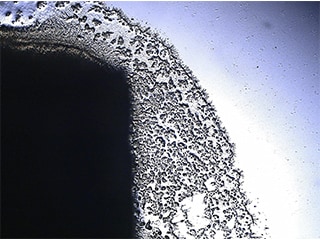
Reasons for Using VHX Series Digital Microscopes
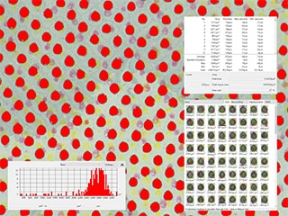
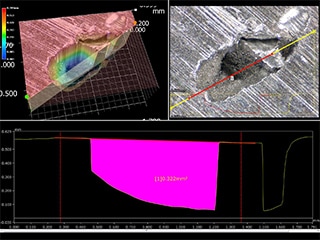
The large depth of field allows the depth composition function to provide observation with the entire image on the screen in focus even at high magnifications
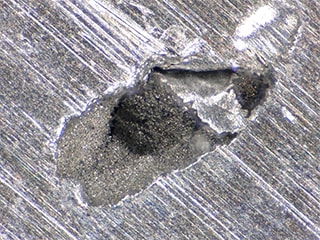
There is no need to cut or polish the target. (Non-destructive observation is possible.)
Being able to select from multiple lighting conditions such as observation with transmitted illumination, ring illumination, coaxial illumination, polarised illumination, and differential interference contrast allows for various applications from biological microscopes to industrial microscopes.
The standard-equipped camera and monitor allow photographs to be captured with the same quality as monitored images


The standard-equipped measuring microscope functions allow for measurement of planes and 3D profiles